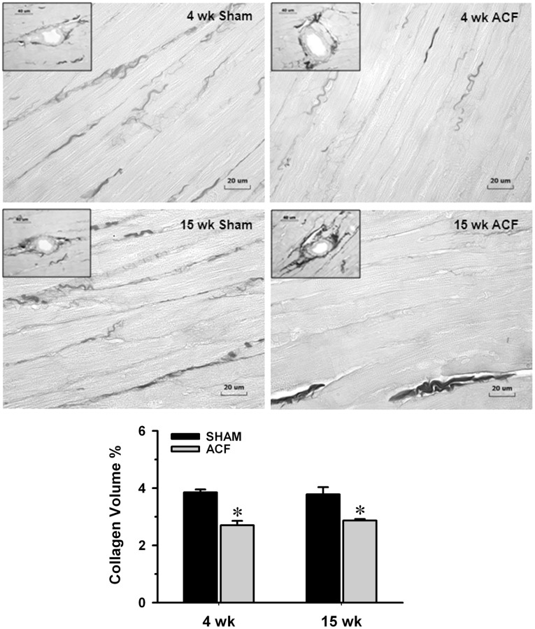
Figure 4. Determination of interstitial collagen volume percent in sham and ACF rats.
Representative images of LV interstitial collagen stained with picric acid sirius red F3BA (PASR) were measured at 4 and 15 wk ACF and their age-matched sham rats. The loss of interstitial collagen (shown as dark collagen fibers excluding perivascular areas) between cardiomyocytes at 4 and 15 wk ACF was reflected by the decrease in collagen volume percent (%). However from insets, when comparing the 4 and 15 wk ACF, there is an obvious increase in perivascular collagen compared to sham in the 15 wk ACF. Panel in the bottom displays quantification of the interstitial collagen at 4 and 15 wk ACF and their age-matched shams. Values are mean±SEM. n = 8–10 in each group. *P<0.05 vs. age-matched shams. Bar scale: 20 µm and 40 µm in insets.

