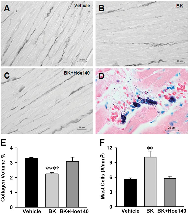
Figure 5. Effect of LV interstitial bradykinin infusion on mast cell infiltration, degranulation and collagen loss.
Interstitial collagen volume percent was decreased 30% by bradykinin infusion group (B) compared to saline infused vehicle control (A) and prevented by Hoe-140 (C). Infusion of bradykinin into LV myocardium increased mast cell number by 2-fold compared to vehicle group and mast cell increases were inhibited by Hoe-140 (F). Representative image (D) demonstrates mast cells and mast cell granules show degranulation after interstitial bradykinin infusion. The number of mast cell was quantitatively determined for the entire LV wall using Giemsa-stained paraffin sections. It produces intense staining (purple) specific for mast cell granules. Panel (E) displays quantification of the interstitial collagen of normal rat with interstitial saline or bradykinin (5 ng/ml) infusion for 24 hrs with and without BK2R antagonist (Hoe-140, 0.5 mg/kg/d given by osmotic infusion pump). Values are mean±SEM. n = 6 in each group. ***P<0.001 vs. Vehicle. † P<0.05 vs. bradykinin infusion plus Hoe 140. **P<0.01 vs. Vehicle and bradykinin infusion plus Hoe-140. Bar scale: 20 ∝m.

