Fig. 3.
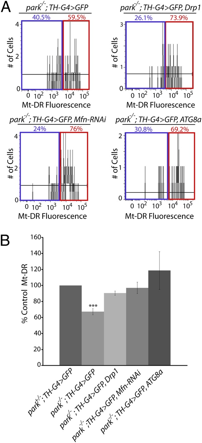
Genetic perturbations that increase mitochondrial fragmentation and turnover rescue the MMP defect of parkin mutants. (A) Neural cultures from parkin-null homozygotes expressing GFP (park−/−; TH-G4 > GFP), from parkin-null homozygotes overexpressing GFP and Drp1 (park−/−; TH-G4 > GFP, Drp1), from parkin-null homozygotes overexpressing GFP and an RNAi construct targeting Mfn (park−/−; TH-G4 > GFP, Mfn-RNAi), or from parkin-null homozygotes overexpressing GFP and ATG8a (park−/−; TH-G4 > GFP, ATG8a) in DA neurons were labeled with Mt-DR and analyzed by flow cytometry. Representative histograms show the percentage of neurons that exhibit Mt-DR fluorescence intensities above and below the cutoff intensity value. (B) Depiction of the mean MMP of DA neurons from animals of the indicated genotypes relative to parkin-null heterozygotes (park+/−; TH-G4 > GFP). The number of biological replicates (n) and total number of cells analyzed (N) for the following genotypes were as follows: park−/−; TH-G4 > GFP (n = 3; N = 299); park−/−; TH-G4 > GFP, Drp1 (n = 3; N = 189); park−/−; TH-G4 > GFP, Mfn-RNAi (n = 3; N = 218); park−/−; TH-G4 > GFP, ATG8a (n = 5; N = 154). ***P < 0.001.

