Fig. 2.
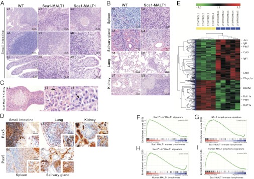
Characterization of human-like MALT lymphomas arising in Sca1-MALT1 mice. (A) Representative hematoxylin–eosin (HE) staining analysis of the mouse lymphomas. A typical lymphoid cell population is present in the small intestine of Sca1-MALT1 mice, infiltrating the lamina propria and the epithelium in small groups, giving rise to lymphoepithelial lesions. (B) Lymphoid tumor cells also infiltrate other tissues. (C) Lymphomas developed in the kidneys showed prominent plasma cells surrounding blood vessels, which were pleomorphic and showed occasional binuclei (marked with arrows). (D) IHC analysis of Pax5 expression in Sca1-MALT1 lymphomas. (E) Gene-expression microarray analysis defined the Sca1-MALT1 lymphoma transcriptional signature. Five Sca1-MALT1 splenic lymphomas and four WT spleen were studied. (F–I) Bioinformatic studies of Sca1-MALT1 murine lymphoma transcriptional signature using GSEA.

