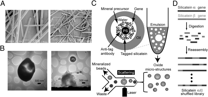
Fig. 1.
Overview of the biomimetic mineralization platform used in this study. (A) SEM images of biosilica spicules (Left) and occluded polymerized silicatein filaments (Right) isolated from the marine sponge T. aurantia. [Reproduced with permission from ref. 29 (Copyright, National Academy of Sciences of the United States)]. (B) TEM images of silicate microstructures formed from biomimetic vesicles, revealing intact bead-silicate composite (Left) and a polystyrene (PS) microbead scaffold released from the fracture of its silicate shell (Right). (C) Polystyrene microbeads coated with silicatein α [by in vitro compartmentalization (20)] are reacted with small metal-containing precursors in water-in-oil emulsions, yielding mineral composites such as those shown in B, which are then isolated from nonmineralized polymer beads by flow sorting, with light scattering used to identify large beads for sorting. (D) Schematic summary of the gene library used for evolutionary selection experiments. Recombinant genes for two natural isoforms of silicatein were digested and reassembled by DNA shuffling (Materials and Methods) to produce a chimeric library of variant silicateins, which were then screened to identify previously undescribed mineralizing variants via the process summarized in C.

