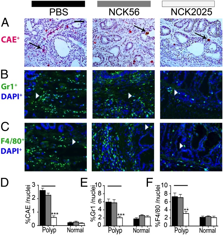
Fig. 3.
Attenuation of intrapolyp inflammation in TS4Cre × cAPClox468 by NCK2025 treatment. (A) CAE-positive mast cells, (B) Gr1+ granulocytes, (C) F4/80+ macrophages, (D) frequencies of intrapolyp CAE+ cells (***P < 0.0001), (E) frequencies of intrapolyp Gr1+ cells (***P < 0.0005), and (F) frequencies of intrapolyp F4/80+ cells (**P < 0.006). (Scale bar: 50 μm.) Black arrows show mast cells, and white arrowheads show F4/80+ or Gr1+ cells for PBS solution-, NCK56-, and NCK2025-treated mice (n = 5 per group). Error bars show SEM. Data were analyzed by two-tailed Student t test, type 2, compared with untreated mice. Sections (5 μm) of frozen colon tissues were stained with CAE-specific antibodies as described previously (13). For immunostaining, primary antibodies used were rat anti-mouse F4/80 (Abcam), biotin anti-mouse Gr1, and hamster anti-mouse CD11c (BD Biosciences); secondary antibodies (DAPI; Invitrogen) were anti-hamster Alexa Fluor 594 and anti-rat Alexa Fluor 488. Tissues were then reacted with streptavidin 488 and DAPI to reveal nuclei. Images were acquired by using a TissueGnostics Tissue/Cell High Throughput Imaging and Analysis System and analyzed by using ImageJ software. Data are representative of three independent experiments.

