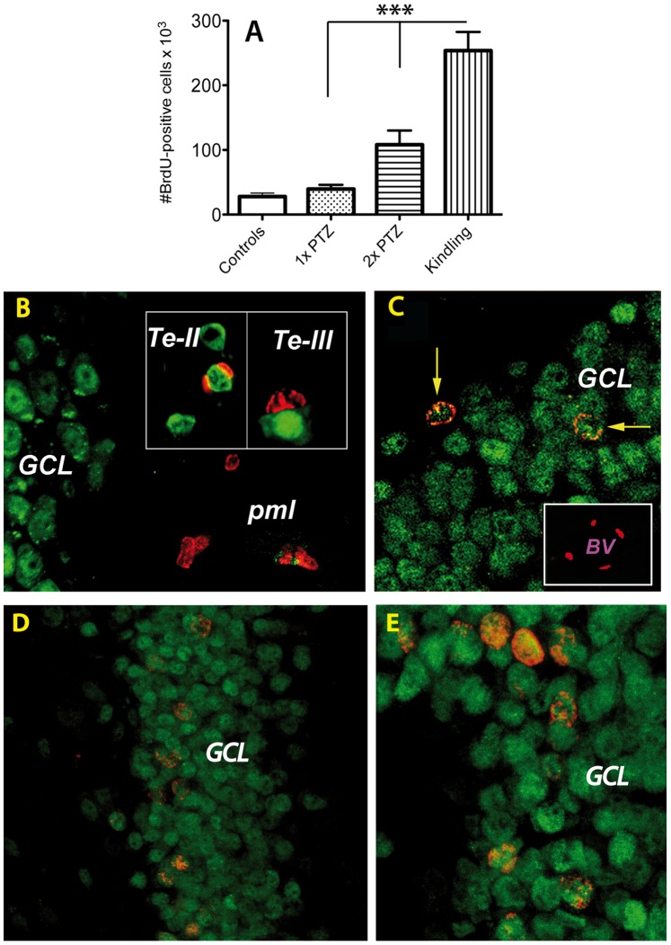
Figure 3. Number and phenotyping of BrdU-positive cells after seizure activity.
Each PTZ treatment led to an accumulation of BrdU+ cells in the dentate gyrus of the kindled rats (9-fold, p = 0.0001; A). (B–E): 3D projections of confocal BrdU(red)/NeuN(green) double-labeled images from PTZ-treated animals. A single episode of seizure activity led to the appearance of BrdU-positive cells in the polymorphic layer that were in a mitosis-like state (B). Occasionally some neurons in layers II and III of the temporal neocortex also displayed BrdU+ cells in close apposition to neurons (B, insets). After 2× PTZ some BrdU-positive cells have differentiated into neurons, particularly in the granule cell layer (C, arrows). In addition, some BrdU-positive nuclei were detected in the walls of large blood vessels (C, inset). The number of double-labeled BrdU(red)/NeuN(green) increased with the number of PTZ injections and reached a maximum in the granule cells layer of kindled animals (D, low power; E, higher power). Abbreviations: Te, temporal neocortex; GCL, granule cell layer; BV, Blood Vessel.

