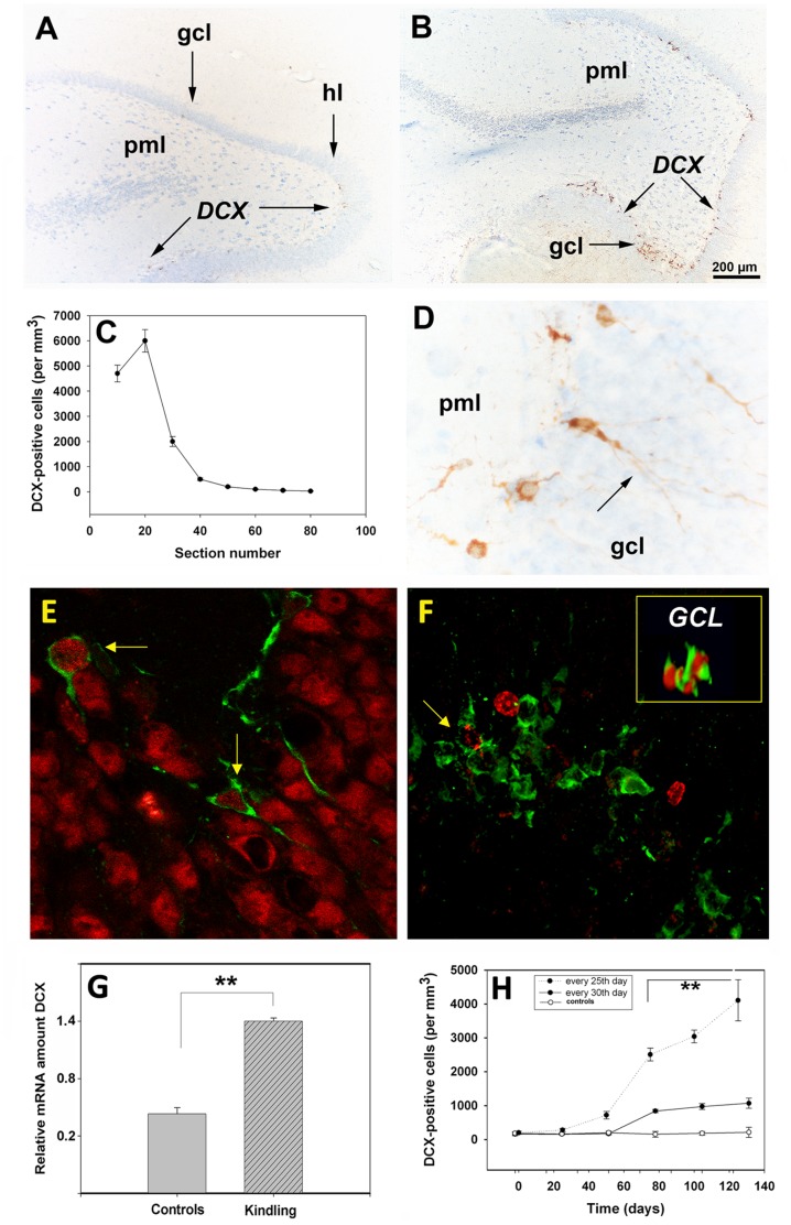
Figure 4. Localization and quantification of DCX in the rat brain during kindling development.
(A, B) Overview of DCX staining in the ventral (A, arrows) and dorsal (B, arrows) hippocampal hilus of the kindled animals. The dorsal hippocampus of kindled animals, was highly significant (p = 0.001) enriched in DCX+-cells (C). Note that the DCX antigens were localized both in cell bodies and extensions penetrating the densely packed granule cell neurons (D, arrows). (E-F): Phenotyping of DCX-cells. After 2× PTZ some DCX+ positive cells (green) in the dorsal hippocampus along the hilar border with the granule cell layer had a NeuN nucleus (red) (E, arrows). In kindled animals some of the DCX (green)/BrdU (red) double-labeled cells had a clonal appearance (F, inset, 3D-image) while other DCX+ cells (green) sometimes displayed a fragmented BrdU-positivity (F, arrow). By quantitative RT-PCR there was a 3-fold increase (p = 0.01) in the relative amount of DCX transcripts in kindled animals over that of controls (G). Note that the number of DCX+ cells also is maximal when PTZ is administered every 25th day (Fig. 4H, filled circles) as opposed to every 30th day (H, open circles). Abbreviations: gcl, granule cell layer; hl, hilus; pml, polymorphic layer. Bars: (A,B), 200 µm; (D), 100 µm.

