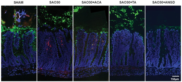
Figure 4. Tissue localization of mucin 2 and trypsin on jejunal sections.
Representative immunostaining of mucin 2 (green), trypsin (red), with nuclei counterstaining (blue) for jejunal sections corresponding to SHAM animals or animals subjected to SAO protocol with luminal inhibition with acarbose (ACA), tranexamic acid (TA) or nafamostat mesilate or without inhibitors (SAO30).

