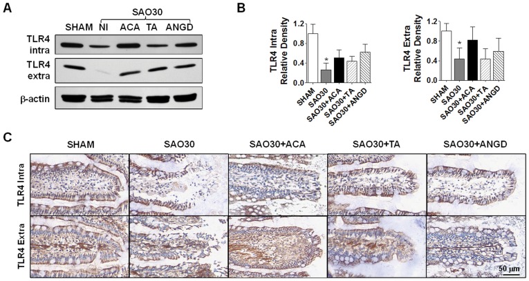
Figure 7. TLR4 is degraded during intestinal ischemia.
Western blot for intra- and extra-cellular domains of TLR4 in jejunal homogenates of SHAM animals or animals subjected to SAO protocol with luminal inhibition with acarbose (ACA), tranexamic acid (TA) or nafamostat mesilate or without (NI) (A) with corresponding density level measurements (B). Immunohistochemistry of intra- and extra-cellular domains of TLR4 (brown) and nuclei counterstaining with hematoxylin (blue) (C). Values are mean±SEM (n = 4)/group *P<0.05 compared to SHAM.

