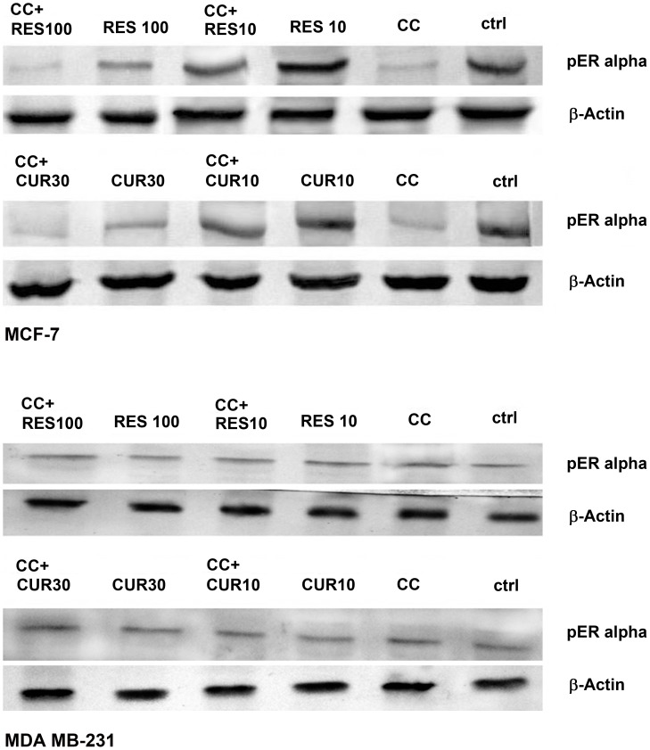
Figure 8. Ser-167 phosphorylation of AF-1 region of ER α.
3×10⁶ MCF-7/MDA MB-231 cells were exposed to CC (10 µM)/polyphenols per se (RES: 10/100 µM and CUR: 10/30 µM) or sensitized with low/high dose polyphenols for 6 h and continued with CC (10 µM) for next 18 h. 50 µg of the whole cell lysate was separated on a 10% SDS-PAGE, probed with respective antisera following transfer to a nitrocellulose membrane with immunoblotting using β-actin as control. Data shown is one of three similar experiments each performed in triplicate.

