
Figure 6. Proteins identified by mass spectrometry are present at inhibitory synapses.
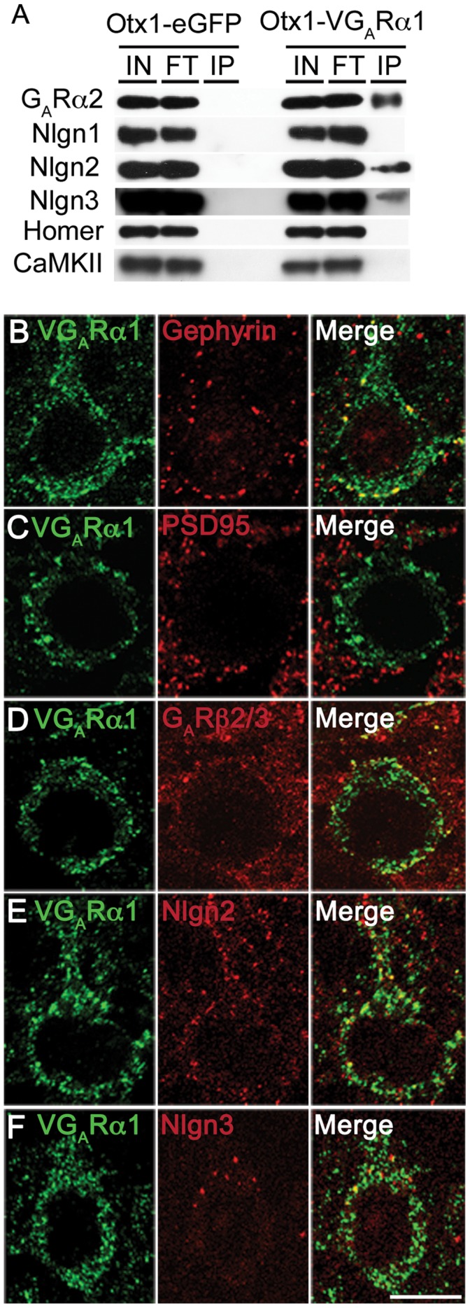
(A) Immunoblotting of several proteins identified by mass spectrometry confirmed their presence in immunopurified inhibitory synapses. GABAARα2, neuroligin2 and neuroligin3 are present, while excitatory markers neuroligin1 and homer are absent from VGABAARα1-tagged inhibitory synapses. The abundant signaling molecule CaMKII is also absent. (B-F) Immunofluorescence studies confirm the colocalization of several proteins identified by mass spectrometry with VGABAARα1. Gephyrin is localized to inhibitory synapses on both the cell soma and axon initial segment (B), while PSD95 is markedly absent (C). GABAARβ2/3 (D), Nlgn2 (E) and Nlgn3 (F) also colocalize with VGABAARα1 in cortical pyramidal neurons. Scale: 10 µm. V: Venus. GAR: GABAA receptor.