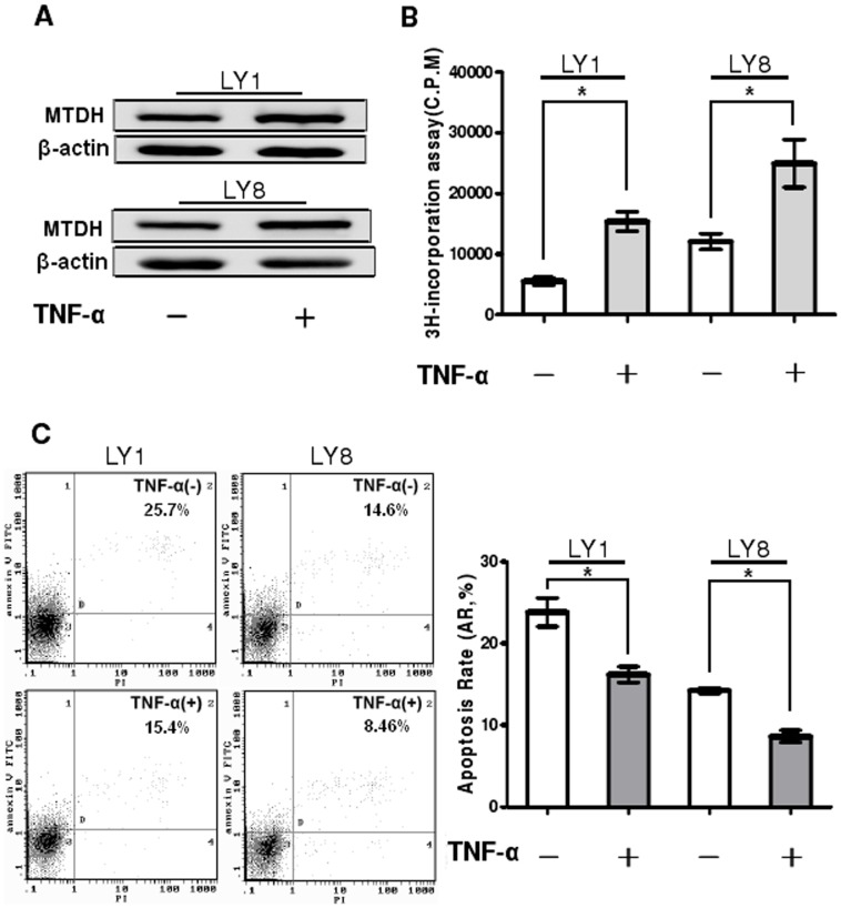
Figure 3. Upregulation of MTDH enhances proliferation and inhibits apoptosis of DLBCL cells.
(A) LY1 and LY8 cells were either untreated or treated with 250 pg/mL of TNF-α for 48 hours. The expression of MTDH protein was analyzed by Western blot. (B) DLBCL cells were treated with TNF-α at the indicated concentration for 48 hours, and cell proliferation was determined by 3H-TdR incorporation assay. Columns indicate mean of triplicate determinations; bars, SD. (C) LY1(left panel) and LY8(right panel) cells were treated with TNF-α (250 pg/mL for 48 hours) and cell apoptosis was detected by flow cytometer. Early apoptotic cells were defined as Annexin-V-FITC-positive, PI-negative cells. Columns indicate mean of triplicate determinations; bars, SD. *p<0.05 versus control.

