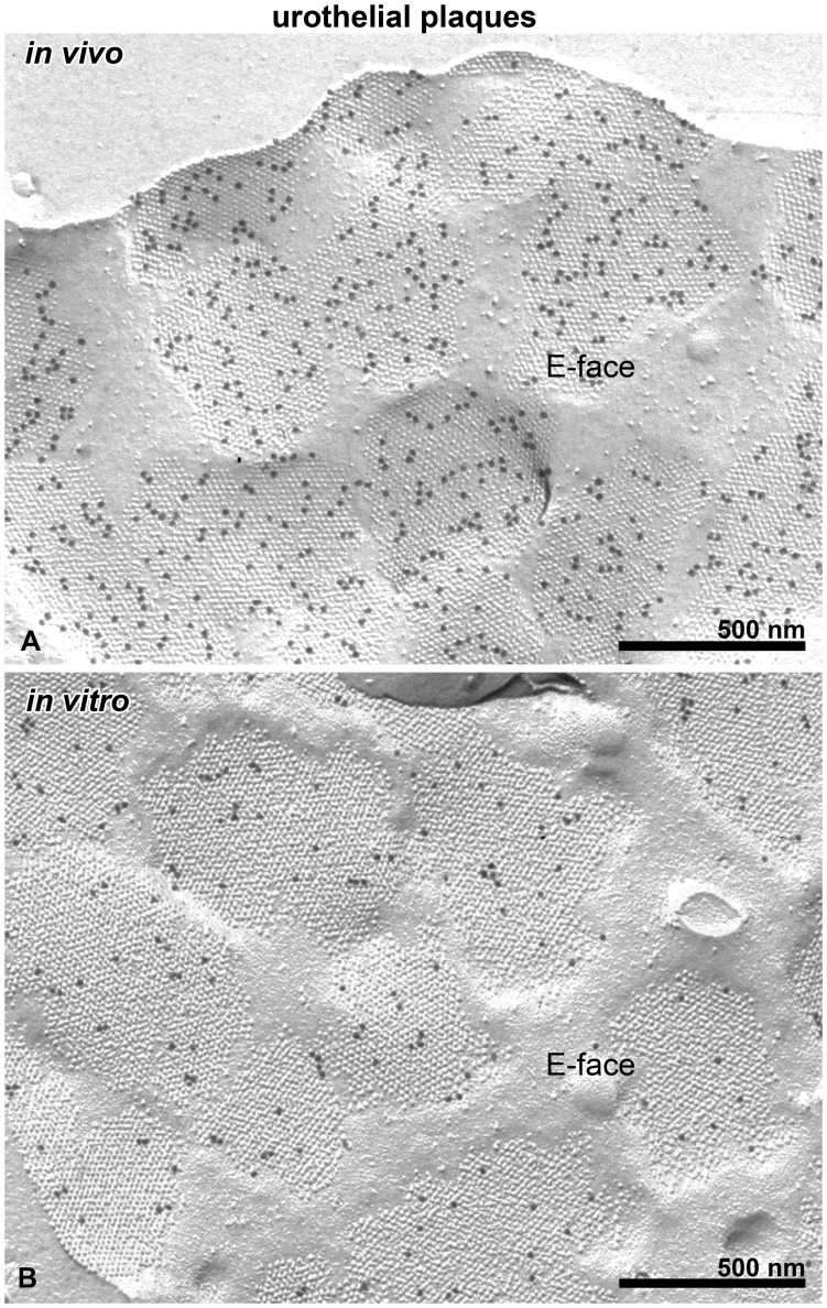
Figure 2. Urothelial plaques are detected in the apical PM of UCs in vivo and in vitro.
Immunogold labelling for uroplakins is seen on the E faces of the apical PM of UCs in vivo (A) and in vitro (B). The maximum calliper diameters and the morphology of urothelial plaques are similar in UCs in vivo and in vitro.

