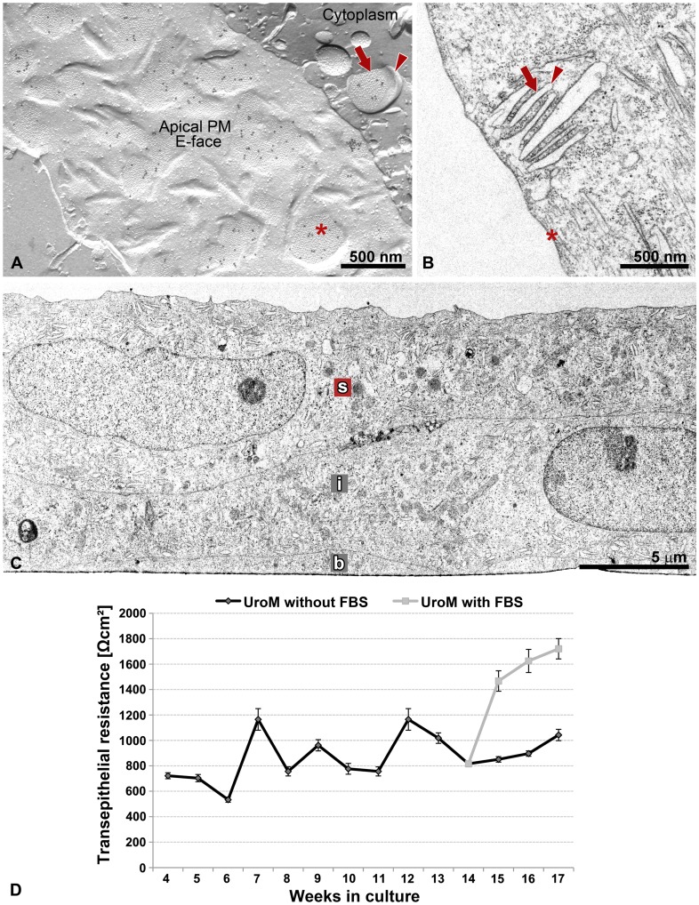
Figure 4. Urothelium in vitro with the ultrastructural and functional properties of native urothelium.
(A) FRIL, (B, C) thin section EM, (D) TER measurements. (A) Image of urothelial plaques in the apical PM and membrane of DFV of UC in vitro prepared by freeze-fracture electron microscopy with immunogold labelling for uroplakins. The uroplakin-positive urothelial plaques in the apical PM (asterisk) and the cytoplasm (arrow) correspond to (B) rigid-looking, concave shaped apical PM structures (asterisk in B) and membranes of the DFVs (arrow in B), respectively, that are visible by thin section EM. Hinge regions are marked by arrowheads in A and B. Note close association of DFVs with the apical PM in A and B. (C) The UCs in vitro, like UCs in vivo, are organized into the urothelium with the basal (b), intermediate (i) and superficial (s) cells. (D) The TER of urothelia grown on porous membrane was measured for 4 months, from week 4 onwards. Each point represents the mean TER (±SE) of urothelia. Urothelia were grown for 14 weeks in the medium UroM without fetal bovine serum (FBS, n = 12), then the two-thirds of urothelia were propagated in the same medium (n = 8) and the other one-third in UroM with 2.5% FBS (n = 4) for an additional 3 weeks. Note the significant increase in the TER of urothelia, which were transferred from the medium UroM without FBS to UroM with FBS.

