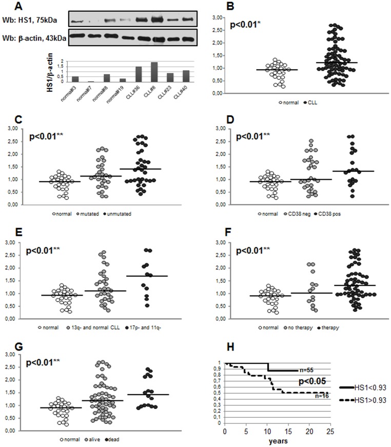
Figure 1. Expression of HS1 protein in CLL B lymphocytes.
The lysates obtained from normal B lymphocytes and leukemic B cells from CLL patients were analyzed by immunostaining with antibody against HS1. Blots were reprobed with anti-β-actin antibody as loading control. Figure 1A is representative of four CLL and four healthy subjects with the respective densitometry of HS1/β-actin ratio. Figure 1B shows HS1/β-actin ratio of 71 CLL patients and 26 normal controls. Data has been normalized putting equal to 1 the ratio calculated in Jurkat cell line. Data obtained were evaluated for their statistical significance with the Student’s t-test (* p<0.01 between normal controls and CLL patients, B) or ANOVA (** p<0.01 between normal vs mutated CLL vs unmutated CLL, C; normal vs CD38 neg CLL vs CD38 pos CLL, D; normal vs 13q- and normal karyotype CLL vs 17p- and 11q- CLL, E; normal vs treated CLL vs untreated CLL, F; normal vs still alive patients vs dead patients, G). Medians are represented by solid lines. Figure 1H represents the overall survival comparison between patients (n = 16) presenting high levels of HS1 (HS1>0.93, dotted line) and patients (n = 54) presenting low levels of HS1 (HS1<0.93, solid line); the difference between curves is statistically significant (p<0.05, Kaplan Meier). 0.93 is the median of HS1 levels in normal controls.

