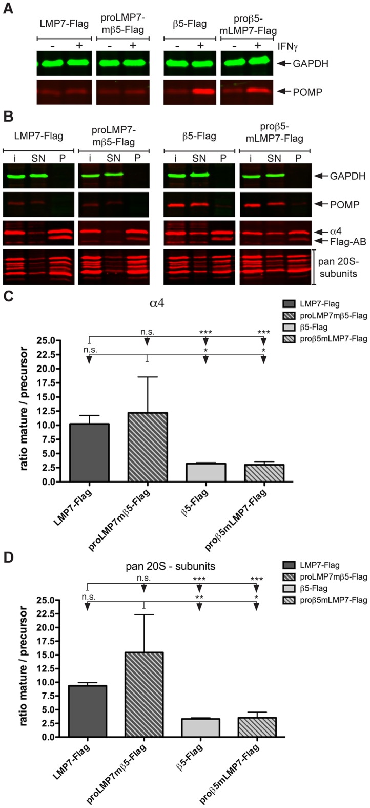
Figure 2. Relative quantification of proteasome maturation in lmp7 − /− Mefs expressing proLMP7- or proβ5-containing subunits.
(A) lmp7−/− Mefs expressing the proLMP7-containing subunits LMP7-Flag and proLMP7mβ5-Flag or the proβ5-containing subunits β5-Flag and proβ5mLMP7-Flag were either left unstimulated or cultured in the presence of 50 U/ml IFNγ for 4 days. The abundance of POMP in the cell lysates of the four cell lines was determined by immunoblot analysis. (B) Immunoprecipitation was performed with anti-Flag M2® agarose using cell lysates of the four different lmp7−/− Mef lines, which were grown in the presence of 50 U/ml IFNγ for 4 days. The abundance of GAPDH, POMP, α4 and pan-20S proteasome subunits (pan 20S subunits) was determined by immunoblot analysis of the input material (i), the supernatants (SN) and precipitates (P) following immunoprecipitation with anti-Flag M2® agarose. (C, D) The ratio mature/precursor resembles the abundance of α4 subunits (C) or pan 20S subunits (D) integrated into mature proteasomes divided by their abundance in precursor proteasomes and was calculated as follows: band intensity of subunit X in the precipitate (P) divided by the band intensity of subunit X in the supernatant (SN). Given values for each cell line are mean values ± standard deviations of at least three independent experiments.

