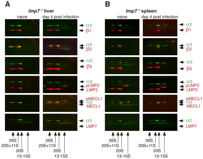
Figure 4. Analysis of proteasome composition in naive and listeria-infected lmp7 − /− mice.
Organ lysates of liver (A) and spleen (B) of naïve and infected lmp7−/− mice were analysed by 2D two-colour fluorescent immunoblot analysis. The mice were infected i.v. with 5×10³ cfu L. monocytogenes and sacrificed 4 days post infection. Organs of three to four mice per group were pooled for the analysis. The different proteasome complexes present in the tissues were first separated by Blue Native-PAGE and subsequently by SDS-PAGE followed by two-colour fluorescent immunoblot analysis. Each membrane was stained for proteasome subunit α3 (green signal) and as it is present in all early to mature complexes in proteasome assembly, α3 serves as a marker for the presence and positions of 13-15S precursor proteasomes, 20S proteasomes and 20S proteasomes + 11S and 19S regulators. Further, membranes were stained for the indicated catalytic β-subunits (red signals) to identify, in which of the indicated proteasome complexes they were integrated.

