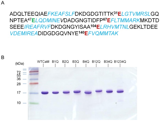
Figure 1. CaM mutant constructs.
A: The sequence of human CaM is shown, using single-letter amino acid residue codes. The mutated amino acid residue in each calcium-binding site is marked in red. Helical regions are shown in Italics and blue color. B: The electrophoretic mobility of wild-type and mutant CaM proteins was analyzed by SDS-PAGE. A 5-μg sample of each CaM construct was loaded in a 15% SDS-PAGE which was run with a standard Laemmli SDS-PAGE buffer in the presence of 1 mM EGTA. Molecular masses of protein standards are shown on the left in kilodaltons (kDa).

