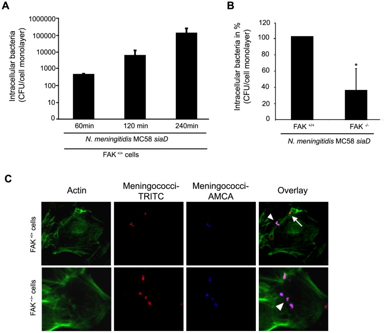
Figure 4. FAK-deficient cells are impaired in their ability to internalize N. meningitidis.
(A) FAK+/+ fibroblasts were infected with MC58 siaD at an MOI of 30 in presence of RPMI cell culture medium, supplemented with 10% human serum (HS). Intracellular bacteria were defined after gentamicin treatment at 60, 120 and 240 min post-infection (p.i.) demonstrating an invasion kinetic similar to 293T cells and HBMEC. The graphs represent mean value ± S.D. of three different independent experiments done in duplicate. * P<0.05. (B) FAK re-expressing (FAk+/+) and FAK-deficient (FAK−/−) fibroblasts were infected with invasive strain MC58 siaD in HS-supplemented RPMI cell culture medium. Intracellular bacteria were estimated at 4 h p.i. by gentamicin protection assays. The graph represents mean value ± S.D. of three different independent experiments done in duplicate. * P<0.05. (C) FAK+/+ and FAK−/− fibroblasts were infected with N. meningitidis strain MC58 siaD for 4 h and analyzed by immunofluorescence microscopy. Extracellular bacteria (arrowhead) stain positive with both TRITC (red fluorescence) and AMCA (blue fluorescence), whereas intracellular bacteria (arrow) are labeled with TRITC only. Cell actin was stained with Alexa Fluor® 488 phalloidin (green fluorescence).

