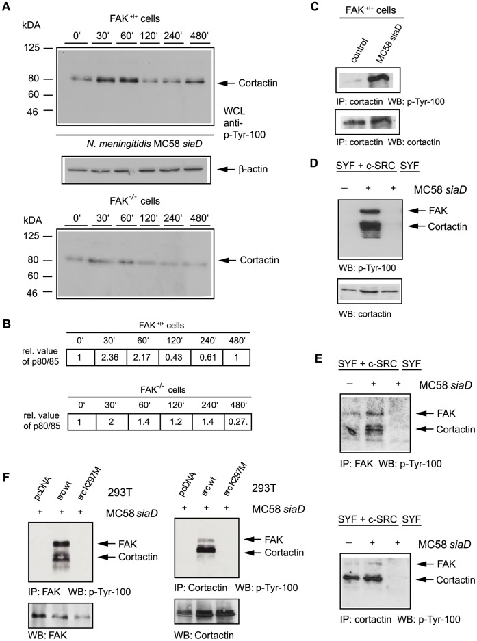
Figure 5. N. meningitidis induced increased tyrosine phosphorylation of cellular proteins.
(A) FAK+/+ cells were serum starved and plated on poly-L-lysine coated dishes. Fibroblasts were left uninfected or infected with N. meningitidis MC58 siaD for a 8 h period and cell lysated were collected at from uninfected control cells and at 30 min, 60 min, 120 min, 240 min and 480 min post-infection and analyzed by Western blotting using the anti-phosphotyrosine antibody p-Tyr-100. (B) The increase of the phosphorylated protein was quantified by densitometric analysis and increase was estimated by comparison to the uninfected control. Densitometric analysis was performed as described in Experimental procedures. Staining of the samples with anti-actin antibody was used as loading control. (C) FAK+/+ cells were infected for 4 h as described above and samples were immunoprecipitated (IP) with an anti-cortactin antibody and immunoprecipitates were analyzed with α-p-Tyr-100 antibody (upper panel). Membranes were stripped and reprobed with polyclonal anti-cortactin antibody (lower panel). (D) Src re-expressing (SYF + c-Src) and c-Src-deficient (SYF) fibroblasts were infected with invasive strain MC58 siaD for 4 h as described above. Cell lysates were collected and analyzed by Western blotting using antibody p-Tyr-100. After stripping membranes were re-probed with polyclonal anti-cortactin antibody (lower panel). (E) SYF + c-Src and SYF cells were infected with strain MC58 siaD for 4 h and samples were immunoprecipitated either with an α-FAK antibody (Fig. 5E, upper panel) or α-cortactin antibody (Fig. 5E, lower panel) and immunoprecipitates were analyzed with α-p-tyr-100 antibody. (F) 293T cells were transfected with the empty control vector (pcDNA), a plasmid encoding wild-type c-Src and a vector encoding the inactive version of Src [Src K297M] to prove that Src kinase activity is required FAK/cortactin phosphorylation. Transfected cells were infected as described above, followed by an IP with an α-FAK antibody or α-cortactin antibody, respectively. Western blot analysis with α-p-tyr-100 demonstrated that Src kinase activity is required FAK/cortactin phosphorylation.

