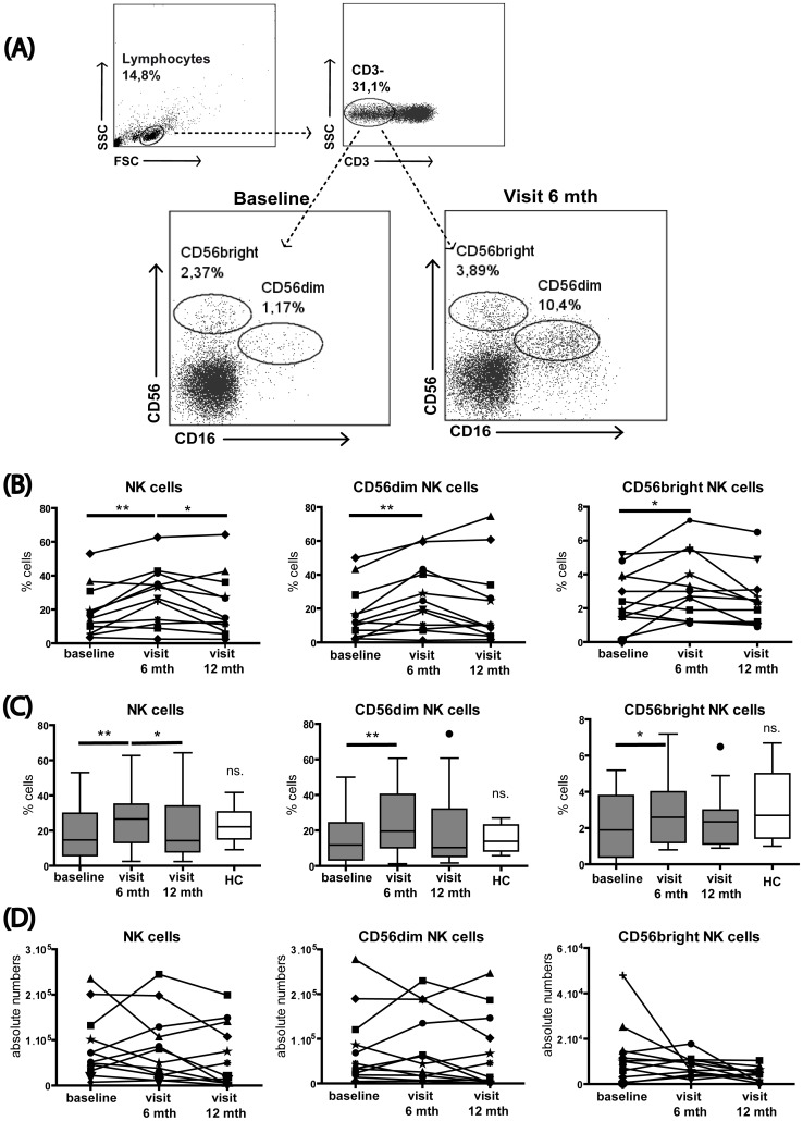
Figure 2. MX treatment promotes NK cell enrichment.
Thawed PBMCs from SPMS patients were stained for NK cells and major subsets using CD56 and CD16. (A) Representative flow cytometry plots show the NK cell gating strategy and MX-induced changes over time in NK cells. (B) Shows the population percentages. After six months of treatment, the NK cell population was significantly enriched and then decreased from six to 12 months of treatment. The CD56dim and CD56bright NK cells subsets were significantly increased after six months of treatment, but no difference was detected from six to 12 months. (C) Frequencies of NK cells and NK cell subsets in SPMS patients before and after MX treatment compared to the frequency observed in matched healthy individuals. (D) Shows absolute counts of NK cells and CD56 subsets: NK cells, CD56dim and CD56bright NK cell subsets remained unchanged over time. *p<0.05; **p<0.01; FSC, forward scatter; SSC, side scatter; mth, months; ns. non significant.

