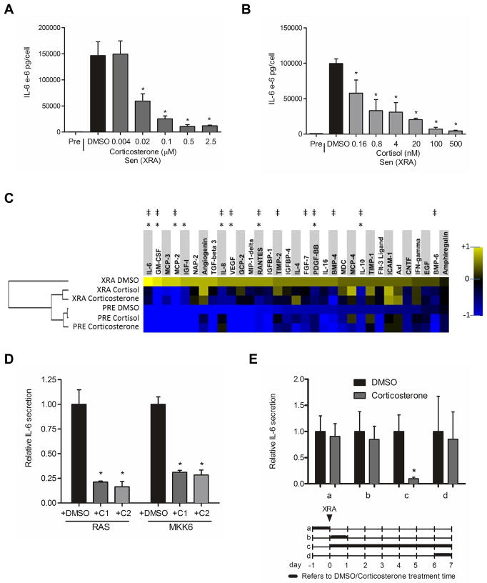
Figure 1. Corticosterone and cortisol partially suppress the SASP.
(A) Senescent (X-irradiated with 10 Gy, (Sen (XRA)) HCA2 fibroblasts were incubated in medium plus 10% serum containing the indicated concentrations of corticosterone or the highest concentration of DMSO (vehicle control). The cells were given corticosterone or DMSO immediately after irradiation and analyzed 6 d later. The cells were washed and incubated in serum-free medium without corticosterone to generate conditioned media. Conditioned media from presenescent (Pre) and control or corticosterone-treated Sen (XRA) cells were analyzed by ELISA for IL-6.
(B) Cells were treated and conditioned media were generated and analyzed as described in (A) except cortisol was used at the indicated concentrations.
(C) Conditioned media were collected from presenescent (PRE) or senescent (XRA) cells that were treated with DMSO, corticosterone (500 nM) or cortisol (100 nM) as described in (A). The conditioned media were analyzed by antibody arrays. We used the average signal from PRE and XRA DMSO cells as the baseline. Signals higher than baseline are yellow; signals lower than baseline are blue. Color intensities represent log2-fold changes from the average value. The hierarchical clustering relationship between samples is shown as a dendrogram (left). *, factors significantly (p<0.05) suppressed by cortisol. ‡, factors significantly (p<0.05) suppressed by corticosterone.
(D) Cells were infected with RAS- or MKK6-expressing lentiviruses. After selection, the cells were given DMSO-, 500 nM corticosterone (C1)- and 100 nM cortisol (C2) for 6 d. Conditioned media were generated as described above and analyzed by ELISA for IL-6. *, factors significantly different from DMSO-treated (p<0.05).
(E) Cells were treated with 500 nM corticosterone for the indicated intervals (a–d, indicated by the thick lines in the lower panel) before or after X-irradiation (XRA, indicated by the arrow). Conditioned media were prepared and analyzed by ELISA for IL-6 (upper panel). *, factors significantly different from DMSO-treated (p<0.05).

