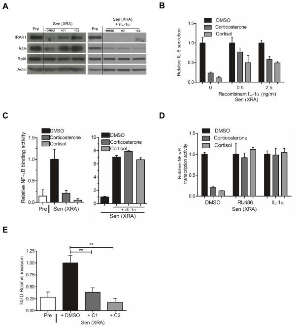
Figure 4. Glucocorticoids impair the IL-1α/NF-κB pathway and suppress the ability of the SASP to induce tumor cell invasion.
(A) Total HCA2 cell lysates were prepared from presenescent (Pre) cells, or senescent cells (Sen (XRA)) cells treated with DMSO-, 500 nM corticosterone (C1)-, or 100 nM cortisol (C2) in the absence (left panel) or presence (right panel) of recombinant IL-1α protein (rIL-1α). The lysates were analyzed by western blotting for IRAK1, IkBα, RelA and actin (control).
(B) After irradiation, Sen (XRA) cells were given DMSO, 500 nM corticosterone or 100 nM cortisol. Six d later, the cells were given recombinant IL-1α protein at the indicated doses in the presence of the glucocorticoids in serum free media. Conditioned media were collected 24 h later and analyzed by ELISA for IL-6.
(C) Nuclear extracts were prepared from Pre cells, and Sen (XRA) cells treated with DMSO, 500 nM corticosterone or 100 nM cortisol in the absence (left panel) or presence (right panel) of recombinant IL-1α protein (rIL-1α), and analyzed for NF-κB DNA binding activity.
(D) Cells were infected with a lentivirus carrying an NF-κB-luciferase reporter construct, irradiated, and allowed to senesce. Immediately after irradiation, cells were treated with DMSO, 500 nM corticosterone or 100 nM cortisol, plus 0.5 μM RU-486 or 2.5 ng/ml IL-1α, as indicated. Seven d after irradiation, cells were trypsinized, counted, lysed and assayed for luciferase activity, which was normalized to cell number.
(E) We prepared conditioned media from presenescent (Pre) cells or senescent cells (Sen (XRA) that had been treated with corticosterone (C1) or cortisol (C2) as described in the legend to Figure 1. The conditioned media were then assayed for ability to stimulate T47D human breast cancer cells to invade a basement membrane, as described in the Experimental Procedures.

