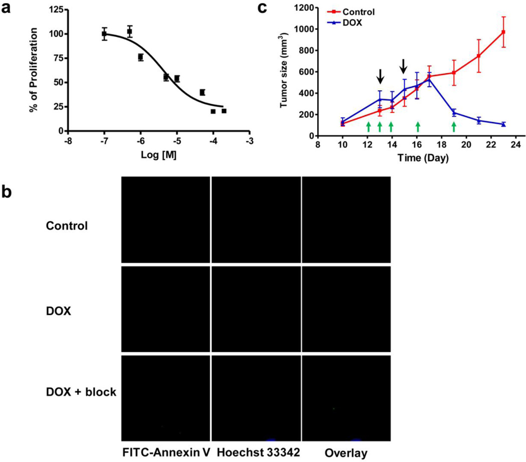
Fig. 1.
Doxorubicin treatment induces apoptosis in UM-SCVC-22B cells ant tumors. a Cytotoxicty of doxorubicin on UM-SCC-22B cells determined by MTT assay. The IC50 is determined as 10 µM after 24 hr incubation. b Fluorescence staining of UM-SCC-22B cells with FITC-Annexin V (green) after treatment with doxorubicin for 24 hr. The cells were co-stained with Hoechst 33342 (blue) for nuclei presentation. c The growth of UM-SCC-22B tumors was effectively inhibited by doxorubicin. The black arrows indicate the time for the two doses of doxorubicin (10 mg/kg for each dose). The green arrows indicate the time for PET imaging studies using 18F-Annexin V.

