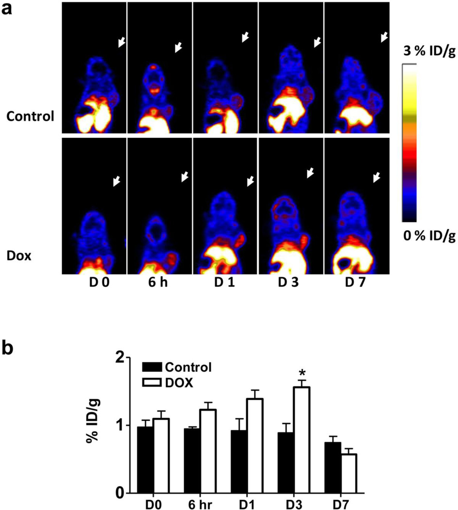
Fig. 3.
18F-Annexin V PET imaging of UM-SCC-22B tumor bearing mice with or without doxorubicin treatment. a Representative decay corrected whole body coronal PET images at different time points after treatment started are shown. 18F-Annexin V (3.7 MBq, 100 µCi) was injected via tail vein and ten-minute static scans were acquired at 1 h after injection. Tumors are indicated by arrows. b Tumor uptake of 18F-Annexin V was quantified from PET scans (n=5). After doxorubicin treatment, tumor uptake increased to a peak at day 3 after treatment started (p < 0.05).

