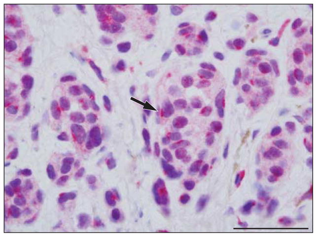
Figure 1.
Benign dermal nevus with soluble adenylyl cyclase (sAC) immunostain and hematoxylineosin costain. The sAC immunostaining pattern (red) is characterized by small dotlike Golgi positivity (arrow) around the nucleus; pannuclear staining is not observed. Weak incomplete granular nuclear staining is sometimes present. Scale bar, 25 μm.

