
Figure 4.
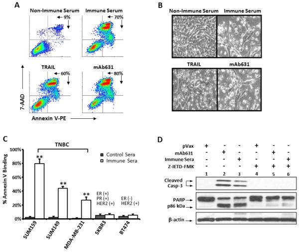

Induction of apoptosis in TNBC cells by hDR5 immune serum. A-B. SUM159 cells were treated for 20 hours with 1% serum from mice vaccinated with pVax1 (non-immune serum) or phDR5 (immune serum) or the hDR5 agonists, mAb631 (5μg/mL) or TRAIL (1 μg/mL). A. Cultured cells were harvested and analyzed by staining with Annexin V and 7-AAD. Percentages represent the number of cells gated as Annexin V/7-AAD-positive cells per total cell count. B. Photomicrographs of representative cell cultures documenting morphological changes consistent with cellular apoptosis. C. The percent Annexin V binding to SUM159 induced by agonist hDR5 antiserum (at 2%) following 20 hr treatment was compared to the indicated TNBC and HER2+/ER+ or HER2+/ER- human breast cancer cell lines (right panel). Statistical analysis was performed using two-tailed Student’s t-tests (**p<0.001; n=4). D. Western blot analysis of SUM159 treated for 5 hours with 2% non-immune (pVax1) or immune (phDR5) serum or 5 μg/mL mAb631 in the absence or presence of 20 μM caspase-8 inhibitor, Z-IETD-FMK as described in Material and Methods. Whole cell lysates were analyzed for cleavage products of caspase-3 (uncleaved caspase-3 not visualized), and PARP; β-actin was used as a loading control. E-F. IgG was purified from immune sera pooled from 5 mice as described in Material and Methods. SUM159 cells were treated for 20 hr with 1% non-immune sera, 1% unpurified pooled immune sera or 1% equivalent purified IgG. Cell apoptosis was measured by staining with Annexin V and 7-AAD (E). Cleavage of PARP was analyzed by Western blot analysis (lower panel) of SUM159 cells treated with the purified IgG, mA631, or non-immune sera. SUM159 growth inhibition was measured by MTT (F) and is displayed as averages of 6 replicates ± SE.