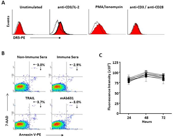
Figure 5.

Resistance of DR5+ activated human T cells to DR5 agonists. A. Peripheral blood T cells were activated with anti-CD3 with IL-2, PMA/ionomycin or anti-CD3/anti-CD28 beads as described in Material and Methods and DR5 surface expression was measured by flow cytometry. B-C. T cells activated with anti-CD3 and IL-2 were analyzed for DR5 agonist-induced apoptosis as assessed by 7-AAD and Annexin V binding (B), as well as Alamar Blue cell survival assay (C); ■ control medium; O non-immune sera (2%); ▲ mAb631 (5 ug/ml); ▽ hDR5 immune sera (2%); □ non-immune sera plus TRAIL (1 ug/ml); ◆ mAb631 plus TRAIL; + hDR5 immune sera (2%) plus TRAIL.
