
Figure 3.
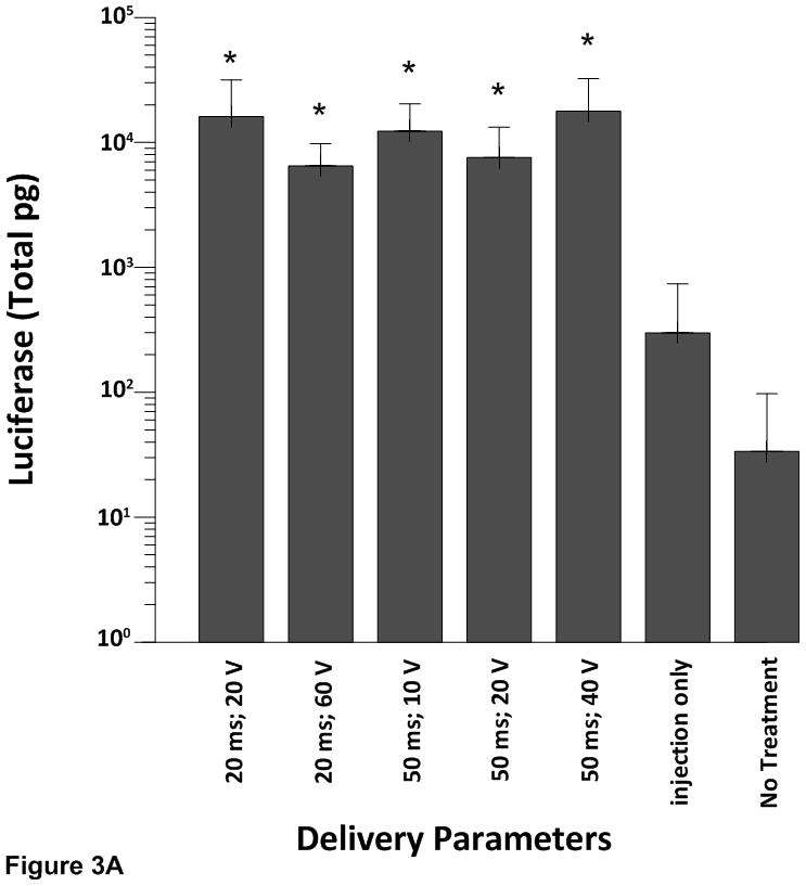
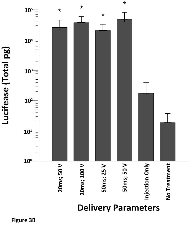
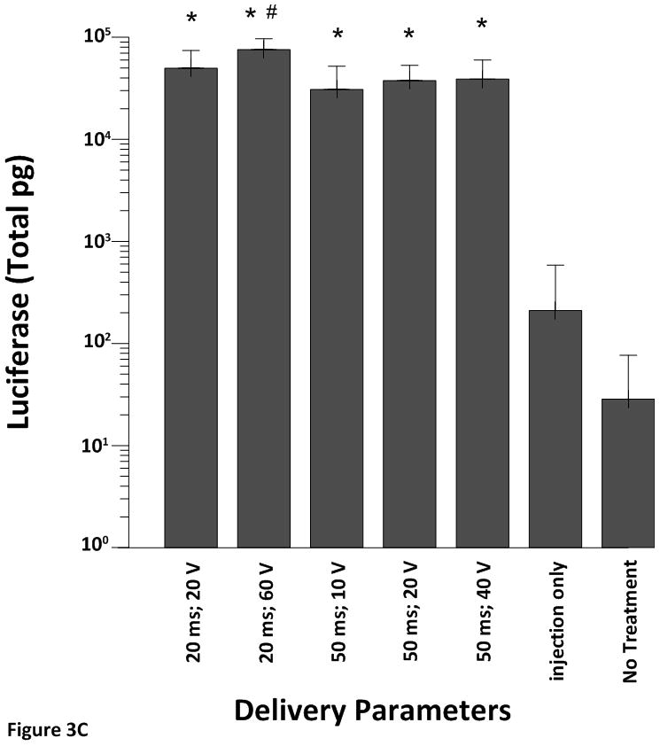
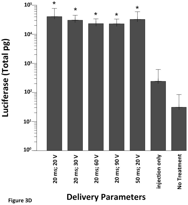
Cardiac expression of luciferase after electroporation-mediated delivery of pLuc. Expression for luciferase is given as the mean total pg ± SD in all figures.
A) Expression levels using the 4 mm penetrating electrode applicator. Injection needle was inserted to a depth of 2.5 mm. B) Expression levels using a non-penetrating electrode.Injection needle was inserted to a depth of 2.5 mm. C) Expression levels using the 7 mm penetrating electrode applicator. Injection needle was inserted to a depth of 3.5 mm. D) Expression levels using the 10 mm penetrating electrode applicator. Injection needle was inserted to a depth of 6.5 mm. Number of sites treated with each electrode delineated in Table 1. An additional 6–10 sites received an injection of pLuc without electroporation (injection only). * p<0.001; # p<0.05.