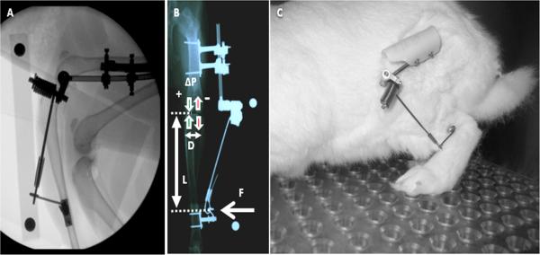
Fig. 1.
A) Fluoroscopy image of a rabbit hind limb showing attachment of the VLD to the bone plates on the lateral femur and tibia and alignment of the axis of rotation of the pivot bearing with the femoral transepicondylar axis. B) AP radiograph illustrating application of varus force to the tibia resulting in increased loading to the medial compartment and decreased loading to the lateral compartment. C) Rabbit with VLD attached and engaged.

