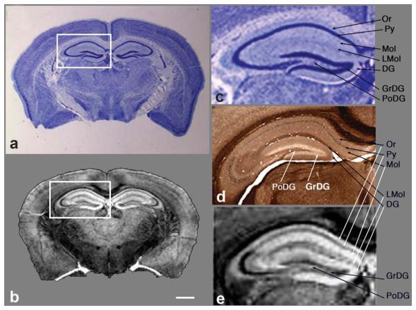
FIG. 5.
Comparison of traditional histology with MR Golgi method. a: Coronal section of a gelatin-embedded mouse brain stained with the thionine/Nissl procedure and (b) corresponding coronal slice from the 3D MR image after chromate plus Gd-HP-DO3A treatment at the level of the hippocampus (boxed region). An expanded view of the hippocampus comparing thionine/Nissl stain (c), peroxidase/diaminobenzidine stain (d), and MRM (e) shows the high level of detail in the MRM image, with excellent correspondence to histologic detail. Note the detailed layers of the hippocampus are easily identified (b,e). Or, stratum oriens; sp, stratum pyramidale; Mol, molecular layer; LMol, stratum lacunosum moleculare; DG, dentate gyrus; sg, granule cell layer; PoDG, polymorphic layer. MRI: 5-mM Gd-HP-DO3A plus 3% K2(Cr2O7). Scale bar = 1mm.

