
Figure 2.
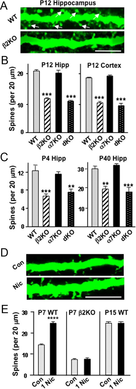
Role of β2*-nAChRs in supporting dendritic spines in vivo. A, Images showing dendrites of cells labeled by intracranially injecting lenti-GFP into WT (top) or β2KO (bottom) mice on P1–P2 and imaging fixed slices on P12 (arrows indicate examples of spines). Scale bar, 5 μm. B, Spine counts per 20 μm dendritic length of WT and KO pyramidal neurons in the CA1 (left, hippocampus) and layer 5/6 pyramidal neurons in the visual cortex (right, cortex). C, Spine counts along sindbis–GFP dendrites of CA1 pyramidal neurons at P4 or P40 (5–10 cells per animal; 3–5 animals per genotype). D, Images of dendrites from P7 Thy-1M-GFP mouse pups, which received stereotaxic intracranial hippocampal injections of PBS (Con) or 1 μm nicotine (Nic) and were perfusion fixed 1 h later. Scale bar, 10 μm. E, Quantification of spine numbers on CA1 pyramidal neuron apical dendrites for P7 WT Thy-1M-GFP, P7 β2KO (sindbis–GFP-labeled), and P15 WT Thy-1M-GFP mice given injections of nicotine (n = 3–5 animals, 4 cells per animal). Hipp, Hippocampus.