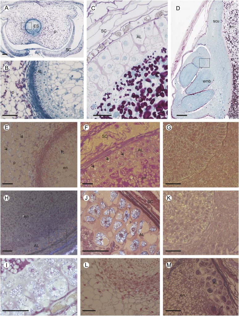
Figure 5.
Morphological and structural observations of developing barley grain, and histological localization of MT proteins. A, General view of a transverse section of developing grain at 7 dap showing the maternal pericarp surrounding the central embryo sac. Polysaccharides and soluble proteins were stained by periodic acid-Schiff (dark pink) and naphthol blue-black (blue). B, Magnification of the 7-dap-old embryo sac wall including the developing aleurone layer between the young endosperm (inside) and the starch-loaded parenchyma cells of the pericarp (outside). C, Median transverse section of a grain at 21 dap showing the aleurone layer and starch grains in the endosperm. D, Longitudinal section of a mature grain including the embryo. The square in D indicates the area corresponding to G and K. E to K, Histological immunolocalization of MT3 and MT4 in developing grain transverse sections stained by fuchsin and examined under bright-field epipolarized light. The silver-enhanced gold particles used for the immunolocalization appear as a bright yellow color. Immunolocalization of MT3 (E–G) and MT4 (H–K) in 7-dap (E), 21-dap (F, H, and I), and mature (G, J, and K) barley grains is shown. MT3 was located in the maternal pericarp, the aleurone cell layer, and the embryo, while MT4 was detected in grains at 21 dap in the aleurone layer and the growing cells of the embryo. L and M, Examples of control sections of grain treated with MT3 (7-dap-old grain; L) or MT4 (21-dap-old grain; M) preimmune serum. AL, Aleurone cell layer; emb, embryo; en, endosperm; ES, embryo sac; P, pericarp; SC, seed coat; scu, scutellum. Bars = 100 µm.

