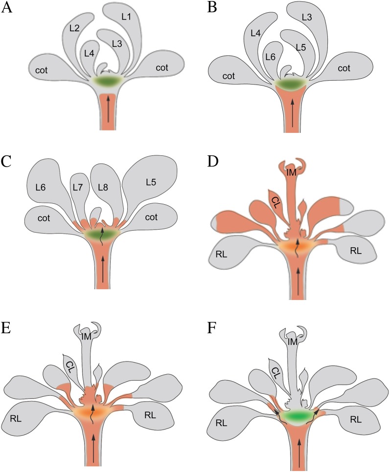
Figure 11.
A simplified model of RtSS. A, A silencing front moves through the hypocotyl cell to cell involving a signal-amplifying mechanism from root to shoot. The red color denotes the silencing front. The green ellipse denotes a tissue domain within the cotyledon node zone. B, When the silencing front reaches the HEJ, just below this tissue domain, the rate of silencing slows. C and D, Once the front gets through/around this barrier (C), the silencing signal moves to the meristem and causes the silencing of all subsequent lateral organs (D). The silencing front can also move into the petioles of older existing leaves and penetrates the lower parts of younger leaves with primary (more open) plasmodesmata. Leaf expansion reveals bizonal silencing in these leaves (D). E, If the silencing signal is generated later, the silencing front can also move through the barrier zone, but it cannot cause silencing of the inflorescence meristem due to the lower rate of cell-to-cell movement. F, The silencing front can be stopped at the barrier zone by the actin stabilizer TIBA, but cell-to-cell movement in other cell types or tissues cannot be stopped. CL, Cauline leaf; cot, cotyledon; IM, inflorescence meristem; L1 to L8, leaf numbers 1 to 8; RL, rosette leaf.

