
Figure 4. The Corticospinal Tract (CST).
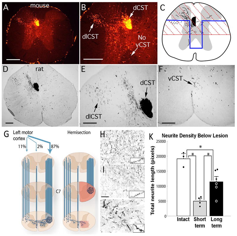
(A, B) Corticospinal axons in a mouse are traced here by making placing four injections of mini-ruby BDA into the right sensorimotor cortex. CST axons from the right hemisphere are labeled. In mice, CST axons descend primarily in two tracts: the dorsal CST, in the ventral part of the dorsal column (dCST), and the dorsolateral CST (dlCST) in the dorsal part of the lateral column. Note the absence of labeled axons in the ventral column on the right side; in rats, some axons are present in this region. Note also the presence of a few labeled axons in the dorsal column on the right in the same location as the dCST. This illustrates the fact that there are a small number of axons that descend in the spinal cord ipsilateral to the cortex of origin. In this case, labeled CST axon arbors are found mainly in the ventral horn gray matter. The distribution of axonal arbors depends on whether the injections target mainly the sensory vs. motor divisions of the sensorimotor cortex. Injections that mainly target the primary motor cortex preferentially label CST axon arbors in the ventral horn whereas injections that mainly target the sensory cortex preferentially label CST axon arbors in the dorsal horn. (C) Schematic illustration of partial transection lesions that are commonly used for assessing CST regeneration in mice and rats. The red region denotes a dorsal hemisection; the blue region indicates a “T” lesion, which is intended to include the ventral CST in rats (modified from Zheng et al., 2006). (D, E) Corticospinal axons in a rat are traced here by making six injections of mini-ruby BDA into the right sensorimotor cortex. Labels are as in A, B. In this case, labeled axonal arbors are found mainly in the dorsal horn gray matter. (F) higher magnification view of BDA-labeled axons in the ventral column (vCST). Scale bars = 250μm. (G, K) Sprouting of CST axons below a hemisection injury in rhesus monkeys. (G) The schematic on the left illustrates the organization of the CST in rhesus monkeys. Descending CST axons from the left motor cortex are shown. The main component of CST axons descend in the lateral column, as in humans. About 87% descend on the side contralateral to the cortex of origin; 11% descend through the lateral column on the ipsilateral side; the ventral CST contains about 2% of the total number of labeled axons. The schematic on the right illustrates the lesion model, a hemisection at C7, and the CST axons that would survive such lesions. The box indicates the CST arbors in the gray matter that arise from axons decussating across the spinal cord midline. (H) Density of BDA-labeled CST axon arbors in the gray matter in intact monkeys (which includes labeled axons from the main tract and the crossing axons); (I) arbors of surviving crossed CST axons at early time points after a C7 hemisection; (J) arbors of crossed CST axons traced 8 months after a hemisection injury, when extensive compensatory sprouting has occurred. (K) Quantitative assessment of the density of crossed CST axons in the gray matter in intact monkeys, at short intervals after the injury, and at long post-injury intervals. By eight months post-lesion, there is a substantial reconstitution of corticospinal axons below the lesion site, due to sprouting of spared contralateral axons. Panels G-K are from Rosenzweig et al., 2010. Scale bar in “I” = 100μm, and applies to H–J.