
Figure 7. Brainstem Motor Pathways: Rubrospinal, Reticulospinal and Vestibulospinal tracts.
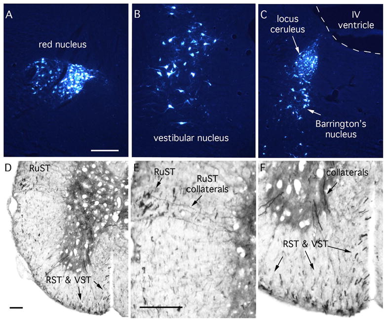
A–C: Retrogradely labeled neurons in the brainstem after injections of true blue into the spinal cord of a mouse. (A) red nucleus; (B) vestibular nucleus; (C) locus ceruleus and Barrington’s nucleus. D–F illustrate BDA labeled axons after a large injection of BDA into the brainstem of a mouse. RuST, rubrospinal axons; RST, reticulospinal axons; VST, vestibulospinal axons. Note collaterals extending from the RuST into the gray matter in E. Collaterals in F are likely from the RST or VST. Scale bars = 250μm.