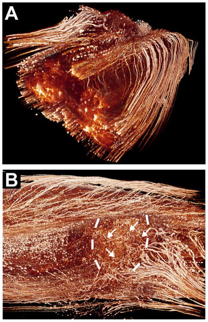
Figure 8. Three Dimensional Imaging of the Unsectioned Spinal Cord.
Using tetrahydrofuran-based methods, the unsectioned adult rat spinal cord can be “cleared,” supporting visualization of the course of individual, fluorescently labeled axons through a spinal cord lesion site (Erturk et al., 2012). This supports tracing the origin and course of individual, lesioned axons. (A) The whole-mounted spinal cord in a 3D representation, showing axons labeled in transgenic M mice (Feng et al., 2000) expressing GFP in sparse neuronal populations (Erturk et al., 2012). (B) Reconstruction of a plane of section from the same spinal cord demonstrating GFP-labeled sensory axons (arrows) approaching and growing within a lesion site (lesion margins indicated by dashed lines). Rats underwent peripheral “conditioning” lesions of the sciatic nerve to enhance regeneration. Caudal is to the right, rostral to the left; the direction of axonal regeneration is right to left. (Courtesy of A Erturk and F Bradke.)

