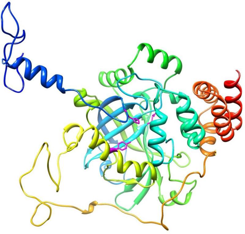
Fig. 3.
A 3D model of the H. vulgaris catalase. Catalase model was generated based on the crystal structure of human erythrocyte catalase (PDB code: 1f4j:A). An individual subunit/monomer of HvCatalase can be seen to possess an eight-stranded antiparallel β-barrel comprising of two four-stranded sheets. The active site heme is surrounded by the β-barrel and α-helices and loops. Conserved catalytic amino acids [His(71), Asn(145), and Tyr(354)] are identified in stick fashion. The figure displayed was drawn using the program Chimera.

