
Fig. 9.
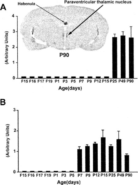
Quantitative analysis of CRF2-mRNA levels in two thalamic nuclei, illustrating the distinct spatiotemporal expression patterns of the receptor in these thalamic regions. Each bar represents means of data derived from a minimum of three animals, with the standard error of the means. The paraventricular thalamic nucleus (A) expresses significant levels of CRF2-mRNA only starting on the fourth postnatal week, while in the ventroposterior lateral and medial thalamic nuclei (VPL and VPM) (B) CRF2-mRNA is observed starting on postnatal day 7 (P7). A strong hybridization signal is observed over the habenula.