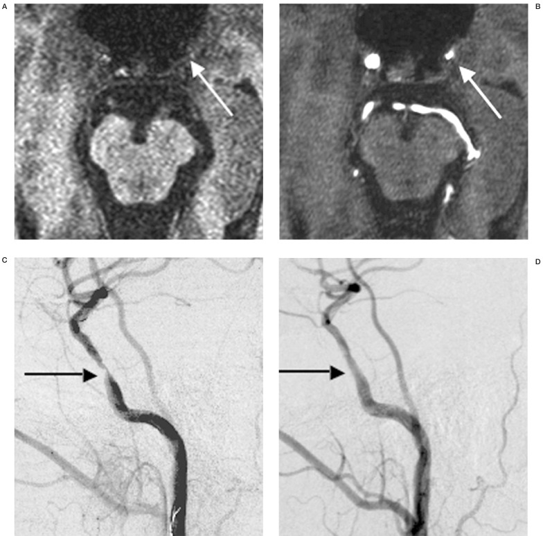
Figure 1.
Case1. A) MPRAGE image demonstrating low intensity plaque of petrous carotid artery stenosis (arrow). B) Source image of 3D-TOF demonstrating petrous carotid artery stenosis (arrow). C) preoperative angiogram demonstrating severe petrous carotid artery stenosis(arrow). D) postoperative angiogram demonstrating good result on PTA(arrow).

