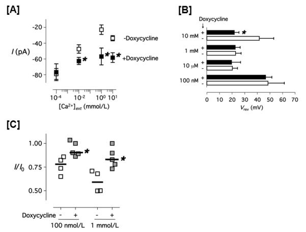
Figure 5. Anomalous mole fraction behavior and calcium-dependent inactivation of the thapsigargin-evoked current in PAECs.

[A] The whole cell transmembrane current values of the thapsigargin-evoked current in PAECs treated with ( ) and without (
 ) doxycycline are shown. Data were collected using the stepwise voltage protocol (see Material and Methods) in 130 mmol/L extracellular Na+ and various extracellular calcium concentrations ([Ca2+]ext). Cells were clamped with the pipette containing 100 nmol/L free Ca2+ and 1 μmol/L thapsigargin, 130 mmol/L extracellular Na+ and the indicated [Ca2+]ext. [B] Reversal potential of the thapsigargin-evoked current in PAECs treated with (closed bar) and without (open bar) doxycycline in the indicated [Ca2+]ext is shown. Data were calculated using a piecewise cubic spline function. [C] The fraction decay of the thapsigargin-evoked whole cell current in PAECs treated with (
 ) and without (
 ) doxycycline, measured for 100 seconds at −80 mV using the stepwise voltage protocol. * P < 0.05 (doxycycline-treated vs. doxycycline non-treated, Student t-test.
