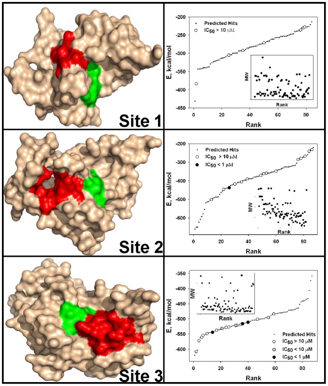
Figure 1. Three docking sites in NS3/4A and VLS of the NCI/DTP compound library.
Left panels, positions of docking sites 1, 2 and 3 in the PDB 3EYD X-ray structure of NS3/4A, surface model. The catalytic triad (His-57, Asp-81, and Ser-139) is green. Docking sites 1, 2 and 3 are red. Right panels, VLS of the 275,000-compound NCI library against docking sites 1, 2 and 3. VLS led to identification of the top 84, 87 and 88 hits, from which 7, 15 and 18 available compounds for sites 1, 2 and 3, respectively, were tested in the NS3/4A inhibitory assays. Compounds were ranked according to their relative binding energy. Black, grey and open circles correspond to the tested compounds with the IC50 values below 1 µM, below 10 µM and above 10 µM, respectively. Predicted (but untested) hits are shown as small back dots. E, relative binding energy. Inset, relations between the molecular weight (MW) and ranking of the ligands.

