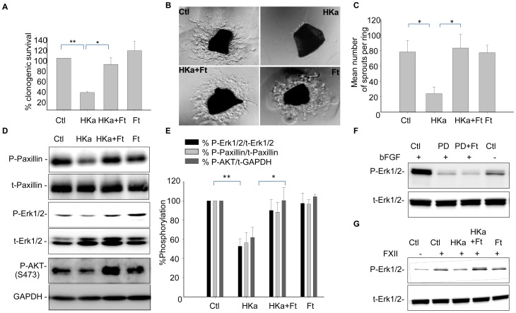
Figure 1. Ferritin restores colony formation, angiogenesis, and phosphorylation of paxillin, Erk1/2, and AKT in HUVECs exposed to HKa.
A. HUVECs were treated with 50 nM HKa with and without 100 nM ferritin in the presence of 20 ng/ml bFGF for 24 hours. Growth medium was replaced and colonies were allowed to grow for 10 days before fixing and staining with crystal violet. Means and standard deviations of 3 independent experiments are shown with *p<0.01; **p<0.002. B. Aortic rings were stimulated with 30 ng/ml VEGF and treated with 100 nM HKa alone or in combination with 200 nM ferritin for 48 hours. Angiogenic sprouts were photographed on day 5. C. The number of sprouts were quantified from three different rings for each condition with *p<0.002. D. HUVECs were treated with 50 nM HKa alone, 100 nM ferritin alone, or co-treated with HKa and ferritin in basal media containing 20 ng/ml bFGF and 10 µM ZnCl2 for 24 hours. Activation of paxillin, Erk and Akt were determined by western blotting using antibodies to phosphorylated (P) and total (T) proteins. E. Band intensities were quantified by densitometry using ImageJ. Means and standard deviations of 3 independent experiments are shown, *p<0.02; **p<0.0003. F. HUVECs were stimulated as in D, and treated with 100 µM PD98059 in presence and absence of 100 nM ferritin. G. Cells were stimulated with 62 nM FXII and treated as described in D.

