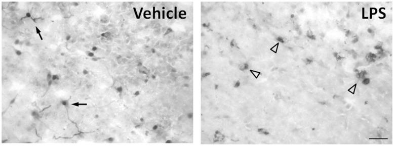
Figure 2. LPS induces microglia activation in vitro.
Representative immunohistochemistry image of frozen pineal sections stained with ED-1 (CD68) antibody. In control tissue, microglial cells are present as small cellular bodies with long branched processes, as expected in a resting or surveillance state (single arrow). LPS changed the morphology of the microglia to cells with larger bodies and no branches, suggesting an activated state (arrow head). n = 3–4 glands. The experiments were repeated twice.

