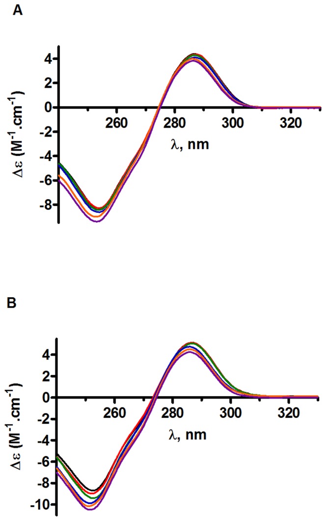
Figure 3. Circular dichroism analysis of oligonucleotides-drug complexes.

Spectra of LTR34 (A) and LTR32 (B) at 10 µM (black) and difference spectra [LTR32/34 (10 µM)+RAL (10 µM, red; 20 µM, green; 40 µM, blue; 60 µM, orange; and 80 µM, purple)−LTR32/34 (10 µM)], in phosphate buffer pH 6, I = 0.05, and MgCl2 5 mM final concentration.
