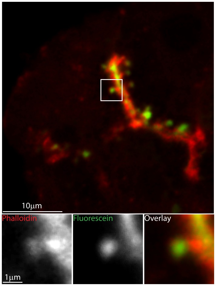
Figure 1. F-actin coats individual fused granules.
Pancreatic tissue fragments were bathed in paraformaldehyde-fixable fluorescein extracellular dye and then stimulated with 1 µM acetylcholine. Each fused granule is then identified by the uptake of fluorescent dye. After fixation, co-staining with phalloidin Alexa-633, shows that each fused granule is coated with F-actin. The upper panel shows a low magnification image, where-as the lower panel shows high magnification images that are enlargements of the boxed region.

