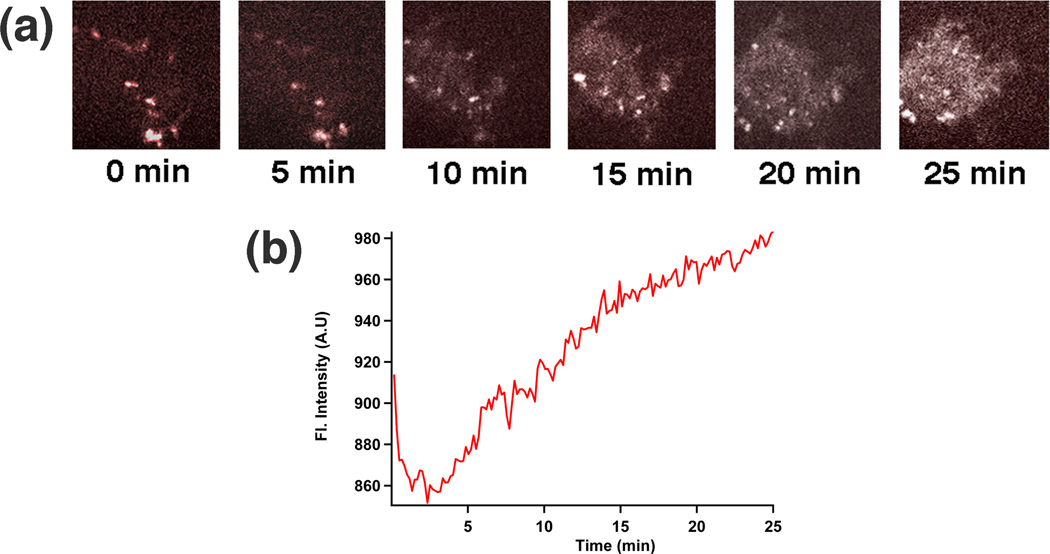
Fig. 5. Real-time imaging of the intracellular delivery event.
(a) Real-time confocal images taken by spinning disk confocal microscopy show the increase of the staining of a cell’s nucleus by PI as a function of time. (b) Plot of the total intensity increase of a cell’s nucleus as a function of time.

