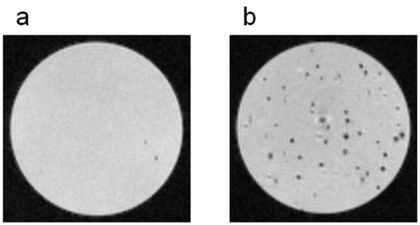
Figure 3.
T2* weighted MRI of single cell suspensions of a) unlabeled MSCs and b) MPIO labeled MSCs in agarose. Cells without internalized iron created minimal signal loss (a), whereas the MSCs labeled with MPIOs induced single, distinctive dark spots throughout the sample (b). Each dark spot corresponds to an MSC with several internalized MPIOs. The MRI images were acquired at a resolution of 50 m.

