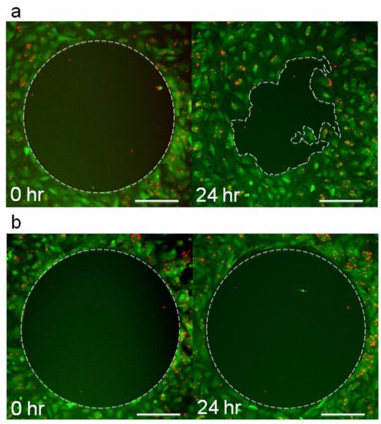
Figure 5.
Fluorescence photomicrographs of chemokinetic migration of MPIO labeled MSCs after 0 and 24 hours of incubation in a) 10% serum containing medium and b) 9L 48 hour tumor conditioned medium. Cells are shown in green and MPIOs in red. Only MSCs in serum containing medium a) showed a chemokinetic response, which resulted in a % closure of ~55 ± 7%. MSCs incubated in serum free medium or any of the tumor conditioned mediums did not migrate into the circular gap, and therefore, did not exhibit a chemokinetic response as shown in b). The scale bars shown are 200 μm.

